Source: Rom J Leg Med [34] 154-161 [2026], DOI: 10.4323/rjlm.2026.154
Kristina Baumjohann, Mark Benecke
Foto: Mark Benecke
Abstract: A man was found lying on the ground in a highway parking lot, covered in blood and severely injured in his head region. Next to him stood another man with blood on his clothes and in his face. The police suspected him of causing the injuries which he denied. We were commissioned to investigate the origin and causes of the bloodstains on the defendant’s clothing and to compare them with his statements and the blood stains on the victim. Experiments with human blood enabled us to approximate the actual sequence of events.
Keywords: forensic biology, bloodstains, blood spatter, stain evidence, experts stain lab.
Foto: Mark Benecke
INTRODUCTION
The examination of bloodstains has been used for over a hundred years to reconstruct the course of crimes and accidents, as well as to verify statements about criminal events [1].
Bloodstains can shed light on moments during the course of the crime, or they can have been created later (by the emergency services, blowflies or similar, see below). It is often possible to obtain precise movement images [2-12].
The shape features of bloodstains and blood drops are influenced, for example, by the type of ground or surface on which the blood has fallen, the height of the bleeding source, the speed of the blood as it fell to the ground and the volume of the drop of blood [13, 14].
Flies can also “cause” bloodstains or alter bloodstains at a later date if the insects regurgitate the blood they have previously ingested or deposit it as feces [3, 15, 16].
Bloodstains can form various patterns on surfaces: For example, the folds in an item of clothing can lead to gaps in the bloodstains and thus provide information about the posture of the wearer when bleeding occurred. Wonder (2001) gives a typical example of this: A woman who has been shot lies on the ground in an outstretched position (Fig. 1) [17].
The perpetrator had claimed that the victim had been standing when the shot was fired. The pattern of blood in the folds of the trousers (shading, fall of the folds) of the dead woman, on the other hand, shows that the blood pattern must have been created in a sitting and not a standing position. Gaps in the bloodstains can provide clues as to the timing of a crime (Fig. 2).
Foto: Mark Benecke
It is sometimes significant that blood which hits dry textile surfaces is (temporarily) not absorbed: according to Wonder (2001), this is said to be due to the fact that the red blood cells cannot migrate through the textile fibers; they must first change their shape and only then can they run around the fibers and penetrate the (fabric) weave [17]. The other blood components (plasma), however, penetrate the fabric without any problems.
When drops of blood come into contact with moist textile surfaces, the membrane of the red blood cells should dissolve immediately, releasing the red blood pigment into the rest of the blood (plasma). As a result, these kinds of bloodstains appear lighter, larger and more irregular than bloodstains on dry textiles [17]. If blood comes into contact with a dry item of clothing and can dry there before it is exposed to moisture or water, the bloodstains are more difficult to remove [17].
The processing of bloodstain samples requires good photographic documentation on site, especially if the presence of blood is considered natural or “self-evident” owing to the existing injuries, as is unfortunately often the case in (car) accidents [18, 19].
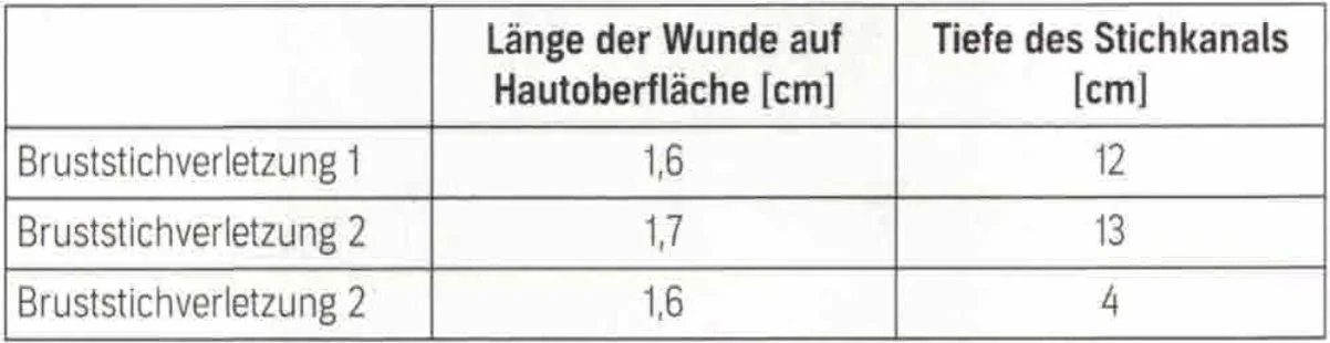
The same applies to serious injuries (cuts to the throat, beating to death, etc.), where the sometimes abundant quantity of bloodstains very well represent the spacetime progression (Fig. 3); although their evaluation requires many measurements of the various bloodstain groups and therefore takes a lot of time. Experiments are often also necessary.
Foto: Mark Benecke
CASE REPORT
The case presented here occurred in the evening at a highway parking lot in Austria. The police found the victim lying on the ground, covered in blood and seriously injured in the head area. The defendant stood next to the victim, his clothes and face covered in blood.
The victim showed signs of blunt and sharp forceful impact including a traumatic brain injury with fracture of the left temporal bone, various hemorrhages in the skull area, bruised areas on the colon with diffuse bleeding, a minor spleen injury, streaky epidermal abrasions on the front of the chest, a bleeding pattern on the chin reminiscent of a shoe print and a cut on the neck, which was medically consistent with the impact of a broken bottle.
Foto: Mark Benecke
The defendant stated that he had nothing to do with the injuries. He had received a hard blow to the head from an unknown person, which initially left him dazed. A short time later, he noticed the victim lying on his back on the ground, covered in blood, and tried to help him. The bloodstains on his body and clothes were created as follows: He had climbed over the injured man’s upper body with his right foot and given him mouth-to-mouth resuscitation. He had laid his right knee on the asphalt at roughly the victim’s hip height and his upper body had been bent over the victim’s upper body. He had probably got the victim’s blood on his face and clothes while he was trying to save him.
Foto: Mark Benecke
Among other things, the defendant had skin abrasions on the front of his knees and a blunt-force trauma on the right side of the back of his head.
Behind the defendant’s vehicle, the police found a mineral water bottle with bloodstains in it. The police suspected that the defendant had wanted to wash the blood from his pants and face with the water. The defendant could not remember this and stated that he had dried or wiped his hands on his sweatpants while trying to help the victim.
The victim and the defendant were intoxicated at the time of the incident (victim: 2.47 g/l blood alcohol = 2.47 percent, defendant: 0.80 mg/l breath alcohol = 1.6 percent).
Among other things, we were tasked with investigating the origin and causes of the bloodstains on the defendant’s clothing. We carried out experiments on the following and used the results to check whether the bloodstains could be reconciled with the defendant’s story:
1. The drying of blood splashes on sweatpants
2. Possible washing off of bloodstains
3. Possible mouth-to-mouth resuscitation
4. Squatting, kneeling
Our experimental case study was based on the following fundamental assumptions:
- The reddish-brown stains visible in the immediate vicinity of the crime in the photos are blood.
- These bloodstains come from the victim.
As the victim of the attack had to be given medical treatment immediately, no genetic material was obtained from under the victim’s fingernails.
MATERIAL AND METHODS
Clothing
In the client’s police forensic laboratory, we examined the trace material (M.B.) with the bloodstains and determined the textile composition of the seized items of clothing.
For the test series, sweatpants were purchased that were sufficiently similar to the original sweatpants in terms of color and fabric composition.
Before the test, the pants were washed at 40 °C with color detergent without fabric softener (Fig. 4). The experiments were carried out with fresh human blood in our forensic laboratory.
Blood sampling
Two series of experiments took place eight days apart: S1 and S2. In S1, the human blood collected in the centrifuge tube was carefully mixed with 2 ml of 1.15 % Na2 EDTA solution per 50 ml of whole blood; while in S 2, the blood obtained in the same way was used without the anticoagulant EDTA (Figs. 5, 6). Before the blood was collected, the experiments had already been set up so that the blood could be used immediately.
Foto: Mark Benecke
RESULTS
Drying of blood splatters on gray sweatpants
The suspect’s sweatpants show splatter marks running from top to bottom in a watery-bloody area of the pants (Figs. 7, 8); the splatter marks have not dissolved in this area (Fig. 9).
This indicates that the suspect’s sweatpants already had dried drops of blood on them when they came into contact with water and/or a blood/water mixture: Small bloodstains remain on the surface of a dry fabric; they become blurred or dissolve on contact with damp surfaces (Fig. 10) (Wonder 2001). This therefore indicates that the victim’s wounds were bleeding profusely when the suspect made contact with the victim; his sweatpants were dry at the time.
If there is fresh blood without water on the pants first, it dries “fixed” very quickly (after two minutes at room temperature) (Fig. 11). This “old” blood is then no longer completely dissolved by moistening with water. Even when kneeling down in a mixture of blood and water with blood-splattered trousers, the original splashes do not completely dissolve (Figs. 12, 13). In both cases the original stains, which had already dried in a short time, remain either permanently or temporarily, depending on the amount of diluting liquid and the drying time.
This shows that drops of a blood-water mixture had also reached the knee area of the suspect’s jogging pants, which were still dry at the time. The blood splatter marks on the back of the sweatpants indicate that the blood had splattered from a point higher than ground level and not from a point flat on the ground (Fig. 14).
The blood did not drip from top to bottom onto the suspect’s trousers by gravity, but by accelerated blood from the sitting or standing victim. It seems unlikely that the blood got onto the suspect’s pants when the injured person coughed it up: The part of the suspect’s pants showing the stains in question would have been shielded during a possible attempted rescue of the injured man who was already lying down.
If the blood got onto the suspect’s trousers while he was standing, it came from splashes from above and not during the attempted rescue of the victim. Even a “shaking off or shaking away with the hands” after the suspect has been rescued cannot be considered to have caused the blood splashes: The small splashes would otherwise have run very strongly or become invisible.
Possible washing off of the suspect vs. dousing of the injured person
The suspect’s statement that he had not significantly washed himself off may be true. Our tests show that instead he knelt in an existing blood-water mixture. In view of the information known to us, this may have been caused by dousing the injured, bleeding person with water.
The suspect’s hands and face were still bleeding when the police arrived: The slightly rounded, rather sharp bloodied edges in the facial hemorrhage are consistent with “slapping one’s own hands in the face” (Figs. 15, 16) and then drying the thin layer of blood on the face. After bleeding and drying the blood on his face, the suspect may (later) have put water on his face; this is suggested by the gaps in the bloodstains around his eyes, which he had kept well closed while he did so.
Possible mouth-to-mouth resuscitation
It appears that mouth-to-mouth resuscitation cannot initially be ruled out in this case. From the point of view of the bloodstains’ investigation, however, the posture of the suspect is significant (Figs. 17-19): It is striking that the crotch of his pants did not have any bloodstains. Mouth-to-mouth resuscitation would have to have been performed entirely from the side (Fig. 19). This evidence is not consistent with the statement he made to the police that he had climbed over the lying man with at least one leg and then performed mouthto- mouth resuscitation: In this position “over” the body of the injured person, the victim’s blood would have reached the crotch area of the trousers.
Squatting, kneeling
Foto: Mark Benecke
A “shading” in the form of an interruption of the blood application can be seen on the lower, rear trouser leg (at the transition between the trousers and the end “cuff” of the sweatpants) of the suspect. This gap in the bloodstain shows that and how the accused was squatting or kneeling at times (Figs. 14, 20). This gap may be (and probably is) a transfer of blood from the suspect’s shoes to his pants: The backs of his shoes have bloodstains on them; the blood probably got onto the hem of his trousers from here (Fig. 21).
This is also shown by our reenactment: The suspect’s statement that he was (in the meantime) bent over the victim with one knee on the ground, the other in the air and both feet on the ground may be correct (Fig. 17). The mere mouth-to-mouth resuscitation of an unexpectedly encountered injured person does not explain (the extent of) the bloodstains on the back of the suspected man’s shoes on their outer sides and soles.
The contact between the two men could not have taken place exclusively on the ground: If they fought with each other, they must also have been exposed to violence or have exercised violence in a standing position. If the suspect had reached the injured man lying on the ground before getting any bloodstains and had just woken up from his faint, neither the missing blood in the crotch of the suspect’s sweatpants nor the blood splash that had previously dried on his gray sweatpants could be explained.
In conclusion, this is a critical case-by-case assessment. We were able to show by means of the bloodstain experiments that:
- the suspect had contact with the person when he was standing or sitting, or in any case not just lying down, as the person was spurting blood or the suspect made swinging movements with his hand or an object
- the injured person bled onto the suspect’s dry pants, which only later got water on them. The defendant’s statements are therefore largely inconsistent with the bloodstain evidence. Conducting the experiments made it possible to verify statements made by the perpetrator in terms of an included/excluded procedure.
The results could not have been achieved through pure reflection.
When conducting an experimental examination, it is always important to use original or near-original objects and trace material from a case and to obtain as many good photos as possible at scale [20]. Experiments represent approximations of the actual course of events and thus shed a light on its measurable parts. It is worth making the effort to carry out an experimental test.
References
→ see .pdf