Insect Traces and the Mummies of Palermo – a Status Report
Insektenspuren und die Mumien von Palermo – ein Statusbericht
Quelle: Entomologie heute 31 (2019): 73–93
KRISTINA BAUMJOHANN & MARK BENECKE
Summary: In an interdisciplinary investigation of the mummies of the Capuchin Monastery in Palermo we made entomological findings with regards to the comparatively poor state of preservation of the deceased, to provide enlightenment about the process of natural mummification, as little is known about how the bodies were handled after death until they were laid out in the basement. Three groups of insects could be found on the bodies that are in accordance with the conditions described and the storage situations in the monastery of Palermo: corpse colonisers, pests and predators. This is our first status report on the topic.
Keywords: Palermo, mummies, insects, forensic entomology, archaeoentomology, feeding traces, decomposition
Zusammenfassung: Bei einer interdisziplinären Untersuchung der Mumien des Kapuzinerklosters in Palermo wurden von uns insektenkundliche Befunde erhoben, um den vergleichsweise schlechten Erhaltungszustand der Verstorbenen wie auch den Vorgang der natürlichen Mumifizierung zu erhellen, da über den Umgang mit den Körpern nach Todeseintritt bis zur Aufbahrung im Keller wenig bekannt ist. An den Leichen konnten drei Insektengruppen gefunden werden, die im Einklang mit den geschilderten Bedingungen und Aufbewahrungssituationen im Kloster von Palermo stehen: Leichenbesiedler, Lästlinge und Räuber. Dies ist unser erster Lagebericht über diese Thematik.
Schlüsselwörter: Palermo, Mumien, Insekten, forensische Entomologie, Archäo-Entomologie, Fraßspuren, Verwesung
Fig. 1: The Capuchin Catacombs of Palermo (Le Catacombe dei Cappuccini). a Main entrance. b Corpses in hallways. c-f Current state of preservation of the deceased (“mummies”).
Abb. 1: Der Mumienkeller von Palermo. a Haupteingang. b Anordnung von Leichen in Gängen. c-f Heutiger Zustand der Leichen.
1. Introduction
The Capuchin Catacombs of Palermo (Le Catacombe dei Cappuccini) are located in the current capital city of Sicily underneath the Capuchin Monastery. It houses what is possibly the largest known collection of spontaneously and anthropogenically mummified mummies (SINEO et al. 2008) in the world (Fig. 1).
The Capuchins — a catholic order — settled here in 1534, after they had formed six years previously from the Franciscans (STRÖBL2007). The vault was initially limited to the cavernous area underneath the Church of Santa Maria della Pace. After around 1599, this space was no longer sufficient for the deceased monks and the area was expanded under the ground. The catacombs were created in this manner and gradually expanded in size (STRÖBL 2007). The absorbency of the volcanic tuff stone evidently caused a large number of the bodies to desiccate in a natural manner, so that the large number of deceased persons in the vault were mummified in a natural manner (WUNN 2007; SINEO et al. 2008).
Fig. 2: The ‘Colatoio’, a room in which the corpses were apparently (temporarily?) prepared. The room has full access for arthropods. Cage door is modern and usually closed. Corpses have not been moved as can be deducted from early photographs of the room.
Abb. 2: Das „Colatoio“, ein Raum, in dem die Leichen offenbar (zeitweise?) hergerichtet wurden. Der Raum ist für Gliedertiere frei zugänglich. Die Gittertür ist normalerweise geschlossen und wurde für uns geöffnet. Lage der Leichen durch Fotos belegbar seit Jahrzehnten unverändert.
Well-ventilated mummification areas that have been excavated in the tuff, so-called colatoi, were used to store the dead on transverse terracotta pipes (Fig. 2). The room was sealed for around 8–12 months. After the rooms were opened, the mummies had been desiccated in a natural manner due to the absorbency of the tuff (STRÖBL 2007; WUNN 2007). The bodies were then laid in the fresh air, rubbed down with vinegar and clothed (SINEO et al. 2008). WUNN (2007) mentioned the dusting off of the bodies of arsenic and chalk, before they were positioned or laid out in the corridors and passageways of the vault. The majority of the deceased were laid out in the vault in open wall niches, others were laying in coffins (Figs 1 b–f, 3 a, b, d). Since the beginning of the 17th century, not only monks, but also benefactors of the order and of the monastery were buried in the vault (AUFDERHEIDE 2003; STRÖBL 2007). From 1783, the catacombs were opened to the general public for burials (STRÖBL 2007). The deceased are arranged in groups in the corridors (the clergy, men, women, children, professional groups such as lawyers etc.) (AUFDERHEIDE 2003). The peace of the deceased was destroyed in 1943 due to a bombing attack and in 1966 due to a fire; damaged water pipelines caused water to get into the vault in the 1980s (STRÖBL 2007). The exact and current number of mummies in the catacombs is unknown (SINEO et al. 2008). The majority of the bodies laid out are skeletonised; only a few bodies still exhibit the remnants of former soft tissue or have been preserved completely (BENECKE 2014; Tab. 1).
Fig. 3: Many corpses are in bad shape. a Virgin, b Mechanical damage due to stabilising wires getting loose. c Stuffing of a mummy. d. Mummified brothers; the formerly much better state of preservation is vaguely visible.
Abb. 3: Heutiger, teils schlechter Zustand der Leichen. a Jungfrau. b Teils mechanischer Zerfall durch Herausrosten der Aufhängung aus der Wand und Zusammenfallen der Leiche. c Ausstopfung einer Leiche bzw. deren Kleidung. d Verstorbenes Brüderpaar; die ehemals gute Erhaltung und Präsentation ist noch schwach zu erahnen.
Inside of the catacombs, different spaces were (and are still) reserved for different socio-economic groups. Priests and monks were distinguished because they lived in different parts of town and also maintained different lifestyles. Whilst the life of monks was strictly regulated (mostly prayer, rest, work inside of the monastary, few hours of other occupations concerning the care of elderly and sick persons), priests were more on duty in their respective parishes. Virgins — i.e. unmarried females with a proper lifestyle — as well as females were placed in a different part of the catacombs, most likely due to the catholic custom to separate women from man, even living persons in churches. Until recently, this habit of separation was common in many catholic churches. Lawyers were considered to be of high social status but may also have had enough money to grease their way into the catacombs — those on display in the catacombs were considered to be honorable persons. The category “males” is likely a mixture of “all remaining social groups”. In archaeological work, it are frequently only fragments of insects that are encountered (GROTE & BENECKE 2001; COURI et al. 2009; HUCHET & GREENBERG 2010; HUCHET et al. 2013). These “witnesses from the past” may provide information about the mummification process (HUCHET 2010). HUCHET (2010) sees information content in this “natural” archive of archaeoentomology that frequently remains undiscovered.
Our archaeoentomological investigations were expanded to include taxonomical and ecological data from forensic entomology that were obtained in succession studies of colleagues (e.g. PAYNE 1965; TRUMBLE & PIENKOWSKI 1979; BUCHELI ET AL. 2009; ANDERSON 2011). Animal cadavers are used to simulate human corpses that are colonised by insects under various different conditions (e.g. indoors vs. open land, water, sun vs. shade, (partially) buried, clothed, burned, day vs. night etc.) (e.g. HASKELL et al. 1989; GREENBERG 1990; DAUTARTES 2009; DE JONG et al. 2011; CABALLERO & LÉON-CORTÉS 2014). Information about early or late corpse colonisers, the location of egg laying, the development of larvae, which traces insects leave behind on corpses, preferred biotopes and living conditions of different groups and species of animals was obtained in this manner (e.g. HWANG & TURNER 2005; DEKEIRSSCHIETER et al. 2009, 2013; MATUSZEWSKI et al. 2013).
Tab. 1: Current state of preservation of the mummies. 1 = Soft tissue of head preserved (fully or partially), no signs of tissue destruction by insects, 2 = tissue loss due to insect activity, 3 = skull skeletonized or only minor pieces of dried tissue visible, 4 = unknown (e.g. hooded by a cowl), 5 = empty pupal shells or fragments of beetles.
Tab: 1: Aktueller Zustand der Mumien. 1 = Weichgewebe des Kopfes (ganz oder teilweise) erhalten, ohne Fraßspuren, 2 = Insektenfraßspuren im Weichgewebe, 3 = skelettiert (auch im Sinne von: kleinste Gewebsstücke ohne Fraßspuren), 4 = nicht erkennbar (bspw. wegen Abdeckung durch Mönchsgewandskapuze), 5 = andere Insektenspuren außer Fraßspuren wie Fliegenpuppen (leeren Hüken) oder Käferfragmente.
The networking of information between forensic and archaeological entomology should be striven for and should be incorporated in the recording of findings in order to prevent misinterpretations and incorrect conclusions. Our one week long interdisciplinary investigation of the mummies of the Capuchin monastery took place in July 2012 at the request of the Head of Capuchin Order of Italy and was organised by the archaeologist JÖRG SCHEIDT. Among other things, we obtained entomological findings on bodies that were exclusively naturally mummified to the best of our knowledge. The exposed faces of some mummies were investigated for entomological traces such as feeding, excrement and fly pupae in order to obtain information about the reasons for the severe damage to the deceased. Only insects that were already dead when found were investigated, no traps for living animals were set or laid (Fig. 3). Traps were checked in another study relating to pest management in the Palermo catacombs (QUERNER et al. 2018); this was not our focus of attention.
2. Methods
2.1. Collection of the entomological traces
The entomological traces were collected using spring steel tweezers and LED torches and they then were transferred into sample containers containing 90% EtOH.
2.2. Investigation and determination of the entomological traces
A binocular stereomicroscope (Leica MZ12.5, maximum 100x), a microscope (Leica DMLED, maximum 1000x) and the corresponding classification literature (OEHLKE 1969; STRESEMANN 1976a, b, 1978; RICHARDS 1977; MOURIER & WINDING 1979; LINDNER 1981; DISNEY 1983; MÜLLER 1986; SMITH 1986; GERSTMEIER 1998, HURKA 2005, KACZOROWSKA & DRABER-MONKO 2009; SCHAEFER 2010) were used for investigation and determination of the entomological traces.
3. Results
Abb. 4: Examples for locations of arthropod remains in nose and mouth openings.
Fig. 4: Lage einiger Entnahmestellen für Arthropodenreste im Mund- und Nasenraum der Mumien.
3.1. Condition of the areas of the bodies investigated
We investigated a total of 667 mummies. Of these 344 mummies were (part)-skeletonised and also exhibited no insect traces on the few wisp-like, dried areas of skin. Insect feeding traces were determined on 240 bodies, 28 additional mummies showed other traces of insects such as fly pupae or beetle feeding (Tab. 1).
Insect parts were found primarily in the nasal cavities; however, they were also found in the oral cavity, the throat and the eye sockets (Fig. 4). In one case, a fly pupa was secured in a sleeve fold.
The faces and hands of the mummies were freely accessible for the investigation. The traces that we secured were located in the faces and facial cavities. The great majority of them were partially skeletonised and covered with leather-like flaps of skin (Figs 1 b, c; 2 a, d; 3 a, b ,d; 4 a, b, d).
3.2. Insect finds
Our samples include fragments, but also animals that have been completely retained of different arthropods (Figs 5, 6):
- Hydrotaea ignava (Harris, 1780) (Diptera: Muscidae)
- Fannia scalaris (Fabricius, 1794) (Diptera: Fanniidae)
- Conicera tibialis Schmitz, 1925 (Diptera: Phoridae)
- Leptocera sp. (Diptera: Sphaeroceridae)
- Necrobiarufipes (DeGeer, 1775) (Coleoptera: Cleridae)
- Gibbium psylloides (Czenpinski, 1778) (Coleoptera: Ptinidae)
- Oryzaephilus surinamensis (Linnaeus, 1758) (Coleoptera: Silvanidae)
- Alysiinae (Hymenoptera: Braconidae)
- Tinea pellionella (Linnaeus, 1758) (Lepidoptera: Tineidae)
- Pseudoscorpions (Arachnida)
3.3. Archaeological-entomological species information
3.3.1. Hydrotaea ignava (Harris, 1780) (Diptera: Muscidae)
The larvae of Hydrotaea ignava live as saprophages; they feed primarily on dead, organic substances. They are frequently found on cadavers, in excrement, dung, kitchen waste or bird’s nests (GREGOR et al. 2002).
H. ignava have also been demonstrated in traps that have been set with cattle faeces (MARTINEZ-SANCHEZ & MARCOS-GARCÍA 2001).
Fig. 5: Various arthropods. a Coleoptera, undetermined. b Conicera tibialis Schmitz, 1925 (length of fragment including eyes: 1 mm). c Gibbium sp. d Arachnidae. e Oryzaephilus surinamensis (Linnaeus, 1758). f Remains of pupal hard shells.
Abb. 5: Verschiedene Arthropoden. a Coleopteren, unbestimmt. b Conicera tibialis Schmitz, 1925 (Länge des Fragments mit Augen 1 mm). c Gibbium sp. d Arachnidae. e Oryzaephilus surinamensis (Linnaeus, 1758). f Puppenhüllen.
In an investigation for the definition of a forest edge, FIEDLER et al. (2008) demonstrated a preference of H. ignava for the edge of a forest area. They were found as one of the dominating species in dead piglets in the early stage of decomposition and as the only Muscidae that were found there during the gas flatulence. A clear preference for the gas flatulence stage was also demonstrated by MATUSZEWSKI et al. (2010) for both the adults and the larvae of H. ignava.
3.3.2. Fannia scalaris (Fabricius, 1794) (Diptera: Fanniidae)
Fanniidae prefer damp habitats (BOUREL et al. 2004). Fannia scalaris is a cosmopolitan that is primarily encountered in open land (SMITH 1986). Indoor areas where F. scalaris is present are mostly laid out simply, for example washing facilities or rubbish puts. Semi-liquid substances such as faeces, in particular pig manure, but also the faeces of other animals and humans provide optimum conditions for development (SMITH 1986; BYRD & CASTNER 2001). This species is also known as the “latrine fly” as a result (SMITH 1986; BYRD & CASTNER 2001). The larvae develop in dung and also in cadavers, bird’s nests, other insects and human corpses, urine-soaked clothing and other similar contaminated materials. In contrast to the species Fannia canicularis, Fannia scalaris does not tend to enter into closed rooms. If the latrine fly is encountered in indoor areas, this may be an indication of unhygienic conditions (BYRD & CASTNER 2001). Representatives of F. scalaris were demonstrated in a study on 22 exhumed bodies buried at approximately two metres depth in the north of France who had been buried between 2 and 29 months beforehand (BOUREL et al. 2004). GAUDRY et al. (2006) also found F. scalaris on lamb carcasses at a depth of 30 cm. This and other species of Fanniidae may cause uro-genital myiasis and may be encountered in the later stages of decomposition (“cheese-like products”) (SMITH 1986).
3.3.3. Conicera tibialis Schmitz, 1925 (Diptera: Phoridae)
Phoridae may be used for the answering and investigation of forensic biological and criminological questions due to their appearance on decomposing organic materials such as human bodies (DISNEY 1983; GREENBERG & WELLS 1998; DISNEY & MANLOVE 2005; MERRIT et al 2007).
If the typical initial colonisers of corpses such as blowflies (Calliphoridae) are prevented from access to dead bodies, then smaller flies such as phorid flies (Phoridae) may profit from this (DISNEY 2005): Due to their small size of approximately 1.5-2.5mm, they can get through gaps, cracks and niches and can gain access to what are, from a human point of view, “closed” room sand buildings. Conicera tibialis (Fig. 5 b) is also called the “coffin fly” (SMITH 1986) and is one of the few species of flies that can regularly be found on bodies in sealed areas or in the earth (DISNEY 1983; BOUREL et al. 2004). Their body size allows them to burrow through earth layers until they find suitable material for colonisation and reproduction (COLYER 1954; LUNDT 1964; BOUREL et al. 2004; DISNEY 2005; MERRIT et al. 2007). They occur in graves mostly with a high number of individuals (BOUREL et al. 2004; DISNEY 2006; MERRIT et al. 2007). Under the earth on the body and in the grave, C. tibialis carries out several complete generation cycles and does not need to return to the (earth) surface for mating (EASTON & SMITH 1970; SMITH 1986; BYRD & CASTNER 2001; BOUREL et al. 2004). This is possible for them for up to five years after a burial (COLYER 1954; MARTIN-VEGA et al. 2011).
In a report of exhumations from Franconia, HOFMANN found phorids, too. They may have been C. tibialis here (HOFMANN 1886; BENECKE 2008). MARTIN-VEGA et al. (2011) reported a case in which numerous C. tibialis were found in a coffin at two metres depth that was exhumed 18 years after the burial. The body on which the flies were breeding exhibited partial grease wax. It remained unclear how long the body had already been colonised in this case however. MERRIT et al. (2007) reported of numerous pupae at different stages of development of C. tibialis in a grave after exhumation. The funeral had already taken place 28 years before.
3.3.4. Leptocera sp. (Diptera: Sphaeroceridae)
In a decomposition study on buried pig cadavers, PAYNE (1965) determined Leptocera sp. in the gas flatulence stage of decompo sition; this has been confirmed by SMITH (1986) and JOHNSON (2012). The Leptocera genus also occurs frequently in cadavers, in dung and in decomposing plant material, among other places; in corpses in particular as soon as the “caseic fermentation” putrefaction liquid is emitted (SMITH 1986). Together with two other species of fly, L. caenosa is one of the dominating species on exhumed bodies; they also appear in connection with faeces, water toilets and also in caves (BOUREL et al. 2004; VANIN & VERNIER 2005).
3.3.5. Necrobia rufipes (De Geer, 1775) (Coleoptera: Cleridae)
The red-legged ham beetle Necrobia rufipes (Fig. 6 c) is one of around 3500 species of the checkered beetle family (CORPORAAL 1950). Members of the genus Necrobia are also called “bone beetles” (HUCHET 2010) and live primarily as a predator of fly maggots (SMITH 1986). Beetles of this genus colonise dead bodies in an advanced stage of decomposition and feed on the bones and skin (SMITH 1986; HUCHET 2010). They can, however, also be found on corpses at earlier points in time, probably in the hunt for fly maggots that are found on the corpse in lower numbers as the process of decomposition progresses (BYRD & CASTNER 2001). Necrobia can be used for the processing of forensic biological questions such as the questions of the time of colonisation (BENECKE 1998; KULSHRESTHA & SATHPATHY 2001).
Necrobia rufipes was demonstrated by HUCHET (2010) on the mummies of Namenkhet Amun. It is directly associated by him with the ancient Egyptians, as N. rufipes is one of the oldest species of insects that has been found on mummies up until now. The previous name was therefore Necrobia mumiarum 1834, as the Oxford entomologist Reverend F. W. Hope was the first to discover it in the stomach of a mummified ibis and described it (HOPE 1834). LEVINSON & LEVINSON (1985) investigated the supply storage and insect species of the granaries and graves of the ancient Egyptians. N. rufipes was found here particularly in the heads of mummies.
3.3.6. Gibbium psylloides (Czenpinski, 1778) (Coleoptera: Ptinidae)
Beetles of the Ptinidae family live in proximity to humans and in their housing. Gibbium psylloides (Fig. 5 c) was found by LEVINSON & LEVINSON (1985) in the grave of Tut-ankh-amun. Adults and juveniles also eat textiles, wool and dead, desiccated insects. ROESLI et al. (2003) demonstrate them in traps that they set in pet shops. The feeding of G. psylloides is stimulated by sugars, vitamins and inorganic salts alongside proteins (EL-SAWAF & EL-SAYES 1978). The beetles reacted most strongly to the protein casein as a food stimulant. HERFS (1958) reported about feeding damage caused by G. psylloides on buttons that were made of a casein-containing plastic mass (“milk- stone” galatith, a duroplastic consisting of casein and formaldehyde). QUERNER et al. (2018) explain the presence of G. psylloides on the mummies of Palermo due to their preference for dry plant material such as straw that was used to stuff the clothing of the mummies in the crypt. In the shrine of The Holy Severin (4th Century CE), however, 15 beetle wings that originated from the ground beetle Nebria salina (Faimaire & Laboulbène, 1854) and the hide beetle Trox scaber (L., 1758/1767) were found for example in the small-scale contents of the grave (bone constituents, dust, clothing fibres and similar) (BENECKE 2011). N. salina is not a corpse coloniser, it may have been interested in other components in the shrine (or it was moved inside during the multiple opening and displays), while T. scaber is often encountered in old cloths, fur, leather, feathers, in bird’s nests and tree cavities. As in the catacombs in Palermo, various different biotopes can also be recognised in close proximity here.
Fig. 6: Some arthropod remains recovered from the mummies and out of coffins. a Lepidoptera (Tinella pelionella Linnaeus, 1758 (?). b Pseudoscorpiones (length 0.5 mm). c Necrobia rufipes (Fabricius, 1781). d Hymenoptera (length 4 mm). e Diplopoda. f Gamasidae.
Abb. 6: Funde von Arthropodenresten an den Leichen (und aus Särgen). a Lepidopteren (Tinella pelionella Linnaeus, 1758 (?). b Pseudoscorpiones (Länge: 0,5 mm). c Necrobia rufipes (Fabricius, 1781). d Hymenoptera (Länge: 4 mm). e Diplopoda. f Gamasidae.
3.3.7. Oryzaephilus surinamensis (Lin- naeus, 1758) (Coleoptera: Silvanidae)
Oryzaephilus surinamensis (Fig. 5 e) is one of the flat grain beetles and is known as the sawtoothed grain beetle. It is distributed throughout the world and lives in proximity to humans and in their housing. O. surina- mensis prefers starchy supplies, in particular of plant origin and grain (SMITH 1986); it happily eats straw and the larvae of other insects. The sawtoothed grain beetle is highly resistant to the cold and can survive the winter in unheated buildings (SMITH 1986).
O.surinamensis has been found multiple times in mummies or in Mediaeval finds (HALL et al. 2000; VALAMOTI & BUCKLAND 1995). Gibbium psylloides was also discovered in the grave of Tut-ankh-amun by LEVINSON & LEVINSON (1985). ROESLI et al. (2003) obtained evidence of this species of beetle in six of eight pet shops investigated.
3.3.8. Alysiinae (Hymenoptera: Braco- nidae)
The sub-family of Alysiinae includes among others Alysia manducator (Panzer, 1799). This wasp is the parasitic species within the Alysiinae that appears most frequently on cadavers. It parasitizes blowflies and their developmental stages (SMITH 1986).
3.3.9. Tinea pellionella (Linnaeus, 1758) (Lepidoptera: Tineidae)
Moths (Lepidoptera) colonise dead bodies in the last phases of decomposition, when the tissue is desiccated and the fermentation processes have been completed (VANIN & HUCHET 2017). If the case-bearing clothes mothTinea pellionella (Fig.6a) is found more frequently on a corpse than skin beetles, then this is an indication that the dead body must have been very dry at the time that it was colonised by T. pellionella (SMITH 1986). Along with dried tissue, hairs are also in their diet (MAZZARELLI et al. 2014). QUERN- ER et al. (2018) also found this case-bearing clothes moth on the mummies of Palermo, but not, however, in such a great number as their relative, the common clothes moth Tineo labisselliella (Hummel, 1823), that primarily infests and eats items of clothing made from animal wool.
3.3.10. Pseudoscorpions (Arachnida)
Pseudoscorpions hunt house dust mites and book lice and live in a predatory manner. HUCHET (2010) was able to determine them on mummies, but was not able to determine the species (Fig. 6 b).
4. Discussion
4.1. Insect finds (general)
If one compares the entomological finds from the mummies of Palermo with those of the find situations of the deceased which have already undergone the processes of active decomposition and the putrefaction-altered and possible desiccated residual tissue is still present, the mummies exhibit relatively few insect traces. Corpses that are advanced in decomposition mostly exhibit a large number of insects at all stages of development directly on the corpse as well as, depending on the situation where they were found, in their immediate vicinity and in the wider surrounding area.
It must be taken into consideration here that our investigations in Palermo took place several hundred years after death occurred. As far as we know, the mummies were regularly re-clothed and relocated over the years and decades. Which (insect) traces have been lost and the condition their bodies exhibited originally is completely unknown.
For our study, that we had to carry out “on sight”, we were only able to look at the heads and in parts the extremities, because we were not allowed to touch the bodies of the mummies. The bodies were mostly no longer available and had been replaced by frames that bore the clothing of the dead that had been filled with straw.
Our results are based on the traces that can still be determined in the tissue of the corpse that have been retained due to the desiccation of the rotten tissue, such as the maggot feeding, or that likely adhere to the period of the active decomposition of the rotten tissue (such as fly pupae or beetle feeding). When evaluating the traces, we therefore only refer to the traces and the condition of the head region that we have discovered
with the mummies: The relative lack of fragments of typical early corpse colonisers such as blowflies (Diptera: Calliphoridae) is apparent. That they did, however, develop on the corpse after death occurred is shown by the typical feeding pattern that they have left behind in the mouth, eyes and nose (Fig. 6). The strong decomposition of tissue is evidence known from casework for its presence on the corpse at an earlier point in time. In this case, the “earlier point in time” reaches from directly after the death occurred until the “active decomposition” stage of decomposition in which the tissue is putrid and mushy. The faces of the mummies exhibit this “active decomposition” in the desiccated condition, that means that they were not conserved in a natural manner immediately after death occurred such that their tissue remained completely. Most of the mummies dried out only after the tissue had decayed and were then placed as mummies in the vault (Figs 1-4). This would also explain the preservation of the typical insect feeding grooves in the mummified soft tissue.
The dead may possibly have been initially “stored temporarily” in open land or similar immediately after death, so that insects had free colonisation access to the corpse. It is unknown how long the period was between death occurring until the “delivery” into the monastery for additional conservation and under what conditions the corpses were transported. The colonisation of dead bodies can take place very quickly and within a few hours after death has occurred. The larvae hatched from the eggs that were laid on the corpse and then developed on the bodies, even if the bodies were already stored in the mummification room. As some of these rooms also have windows, there is the possibility that the windows may have been opened at times, so that blow flies and other insects could get into the rooms in this manner. In what condition the bodies were when removed from the Colatoio room, and if they then were stored once more to “dry” in open land, is unknown. The exact circumstances of the storage of the corpses and the temporal process could not be reconstructed.
There are no noteworthy indications of secondary destruction of the tissue due to animals such as moths, beetles or rodents. On the areas of the bodies that were accessible to us and investigated, we were not able to determine any feeding or gnawing traces in the face or at the extremities that destroyed the mummies at a later point in time after they had been laid out.
The overall poor condition of the mummies and their decay cannot only be traced back to the activity of corpse-associated insects, as these carried out their damaging activities on the skin tissue shortly after death occurred until storage in the vault. Other insects such as beetles occurring later on corpses contributed to the decay of the mummies during storage and when laid out in the vault, because they fed on the hairs, grain sand straw (filling material underneath the clothing as well as on the textiles, e.g. Fig. 3 a, c, d).
Health-endangering amounts of fungi and other micro-organisms were determined in micro-biological and molecular biological investigations of the interior air and walls of the vault, the items of clothing, of samples of the skin, muscles, hair and bones, and the filling materials of the mummies (PINAR et al. 2013). According to PINAR et al. (2013), they are responsible for the ongoing decomposition processes and the destruction on the dead bodies; they classify the high concentration of fungal spores in the interior air of the catacombs as being hazardous to health.
Flies contributed to the decay of the soft tissue before, during and/or directly after storage in the Colatoio room; they do not, however, feed on the dried tissue, textile materials or straw and grain. The species of beetle feeding on and in the materials described as well as the micro-organisms and fungal spores documented continue to be active however and will also continue to decompose the mummies in the future.
4.2. Insect finds, specific
Typical corpse first colonisers were not present in our insect findings in the mummies. Instead other species associated with corpses such as for example the beetle Necrobia rufipes and the flies Conicera tibialis, Fannia scalaris, Hydrotaeaignava and Leptocera sp. were found. Apart from H. ignava, the fly species named are also frequently found in exhumed bodies and in graves and may indicate that these bodies were colonised at a later point in time (COLYER 1954; DISNEY 1983, 2006; BOUREL et al. 2004; MERRITT et al. 2007; MARTIN-VEGA et al. 2011). While N. rufipes and C. tibialis are typical corpse colonisers, H. ignava, F. scalaris and Leptocera sp. may occur both in corpses and in anthropogenic surroundings (BOUREL et al. 2004).
The unhygienic conditions, clothing contaminated with faeces or urine as they may become contaminated shortly after death occurred due to muscle relaxation, may explain the occurrence of H. ignava, F. scalaris and Leptocera sp. (SMITH 1986; BYRD & CASTNER 2001; BOUREL et al. 2004).
The evidence of F. scalaris on the corpses may indicate colonisation in an “open” environment, as this fly species rarely occurs in closed rooms. How “open” the storage facilities for the dead and whether the Colatoio room that have windows in parts can also be included in this is unknown.
N. rufipes and representatives of the braconid wasp subfamily Alysiinae live in a predatory manner and feed on fly maggots among other things. Alysia sp. is one of the species of parasitic Hymenoptera that appears frequently on cadavers (FREDERICKX et al. 2013). It parasitizes blowfly larvae and their pupae. N. rufipes also feeds on blowfly maggots and prefers to live on corpses with dry tissue. According to HUCHET (2010), the occurrence of N. rufipes can be connected temporally to the time that the mummies were wrapped, as this beetle species belongs to the typical corpse fauna of mummies. HUCHET refers here to mummies that were not dried out in a natural manner, but that were instead treated. In a case reported by BYRD & CASTNER (2001), the mummy was first colonised by N. rufipes during the mummification process, at a time when the body had already dried out.
The beetle species Gibbium psylloides and Oryzaephilus surinamensis are typical fellow occupants of human housing. Their occurrence is possibly connected to the clothing being stuffed with straw and grains. The hump beetle G. psylloides also eats textiles and reacts strongly to the protein casein as a food stimulant. While casein is released during autolytic processes (DERNBY 1918), it cannot be excluded that G. psylloides was also attracted by the casein-containing compounds.
The case-bearing clothes moth Tinea pellionella and the pseudoscorpions can indeed be interpreted as accompanying finds, as they may be characteristic of the situation in which they were laid out in the church with floral decoration, straw packing etc. The moth T. pellionella, however, is also found on corpses with very dry tissue (SMITH 1986) and feeds there on skin and hair (MAZZAREL- LI et al. 2014).
QUERNER et al. (2018) also found the case-bearing clothes moth T. pellionella and the beetle G. psylloides on the mummies of the Capuchin vault. Otherwise, their extremely comprehensive variety of species ranges from wood-infesting beetles to textile material eating beetles, moths, cockroaches and so on.
The pseudoscorpions found on the mummies hunt house dust mites and book lice (SMITH 1986). HUCHET (2010) was also able to demonstrate them. Their occurrence on the mummies in connection with dusty straw and grains as well as the dusty clothing is therefore not a surprise.
The insects that we found on the mummies can be roughly divided into three groups: corpse colonisers, nuisance pests and predators.
4.3. Additional insect species that are associated with mummies and graves
The investigations carried out on the mummies of the Capuchin vault by QUERNER et al. (2018) show only a slight consensus with our investigation. In both studies, the species Tinea pellionella and Gibbium psylloides were determined. QUERNER et al. (2018) caught the animals with sticky traps and pheromone traps. Most of the individuals that were collected in this manner feed primarily on wood, textiles, keratin, food and dried plants. They did not, however, determine any animals that can be described as being typical corpse colonisers.
HUCHET & GREENBERG (2010) investigated the insect fauna of a grave of the Moche culture in Peru. They found approximately 200 fly pupae of the genera Calliphoridae, Muscidae and Sarcophagidae. Alongside wing and pupae fragments from the hide beetle Omorgus suberosus (F.) (Coleoptera: Trogidae), there was indirect evidence of parasitic wasps that left their typical hatching holes in the fly larvae of the flesh fly (Sarcophagidae).
In an investigation in Egypt that was also published in 2010 by HUCHET (2010), blowflies were also found on a mummy. The species that dominated here, Chrysomyia albiceps (Wiedemann,1819), prefers larger bodies as their larvae require uninterrupted access to nutrition for development.
Fig. 7: Beetles found by THOMAS JOSEPH PETTIGREW (1791–1865, known as “Mummy” PETTIGREW), a surgeon and antiquarian in London, who was an expert on ancient Egyptian mummies. Plate V from his book „A History of Egyptian mummies” (1834).
Abb. 7: Von dem Chirurgen und Antiquar THOMAS JOSEPH PETTIGREW (1791–1865, bekannt als „Mummy PETTIGREW“) aus London an ägyptischen Mumien bei „Auswickelungen” angetroffene Käfer; hier Tafel 5 aus seinem Buch „A History of Egyptian mummies” (1834).
Skin beetles (Coleoptera: Dermestidae) are frequent visitors to corpses that are either partially or completely desiccated (PETTIGREW 1834, see Fig. 7; KULSHRESTHA & SATH-PATHY 2001; SCHROEDER et al. 2002; CHARABIDZE et al. 2014). They colonise corpses in the open air (subaerial) as well as inside of housholds and feed on keratin-containing materials (e.g. HUCHET 2014). Dermestids were, however, also observed on freshly dead tissue in our own decomposition studies (BAUMJOHANN & BENECKE 2019). As beetles have biting mouth parts, they are suitable for the later stages of decomposition from a purely morphological point of view, as the corpse tissue is mostly firm, hard and dry. Flies with their licking-sucking mouth parts can find neither liquid substances nor food intake and their offspring, the maggots cannot feed from it either. Fly larvae require soft tissue that they can ingest and scrape with their mouth hooks. PETTIGREW found “Dermestes vulpinus”, “D. pollinctus”, “D. Roei” [sic!] and “D. elongatus” (determined by Rev. F. W. HOPE) on an mummy acquired by a Mr. BRODIE (PETTIGREW 1834, p. 55).
Another corpse-loving beetle is the hide beetle Omorgus suberosus (Fabricius, 1775) that was found by HUCHET & GREENBERG in an additional investigation (HUCHET & GREENBERG 2010). It colonises dead bodies primarily in the last stage of decomposition and feeds from the keratin-containing ma- terials such as hair, ligament, skin and bone (PALESTRINI et al. 1992). We were not able to find any skin beetles or hide beetles on the corpses, in their clothing or in the near surroundings. There was neither the typical beetle feeding of skin beetles nor traces on the bones that indicate this beetle.
In the same study from 2010, HUCHET & GREENBERG found Muscidae pupae of Ophyra aenescens (Wiedemann, 1830) and Synthesiomyia nudiseta (Van der Wulp, 1883). Both species live associated with corpses, whereby O. aenescens is frequently found on buried corpses (BOUREL et al. 2004). O. aenescens and S. nudiseta colonise dead bodies at a later point in time: O. aenescens primarily in the active putrefaction stage (BYRD & CASTNER 2001; TURCHETTO & VANIN 2004) and S. nudiseta after the first wave of colonisation (SKIDMORE 1984). The pupae of flesh flies (Sarcophagidae) were also found in the grave of the dead (HUCHET & GREENBERG 2010); the species could not be determined in more detail. A large portion of these pupae were infested by parasitic wasps (possibly Pteromalidae or Chalcidae); their shells exhibited the characteristic circular hatching holes of the wasp’s offspring.
In Italy, flies of the genera Calliphora and Lucilia are typical initial corpse colonisers (VANIN et al. 2008, 2011; BONACCI et al. 2009, 2017). In Peru, in contrast, Cochliomyia macellaria (Fabricius, 1775), whose larvae were found in a grave, is the most frequent species of blowfly on cadavers (BAUMGARTNER & GREENBERG 1985; IANNACONE 2003). Together with the blowfly Compsomyiops verena (MELO 1968), which was also found in pupae shells in the same investigation, these two species were probably among the first colonisers of the bodies (HUCHET 2010). Blowflies were not able to be determined directly in Palermo. The feeding traces of their larvae in the mummified rotten tissue suggests that they were present, however. Which blowflies left their traces here is unknown. Pupae and their fragments could not be determined. In contrast to the studies on mummies and graves which have already been mentioned, the corpses here have hardly been moved or had their position changed, or were not moved at all after their death, so the migrated and pupated fly larvae were able to be found as pupae shells.
The numerous feeding traces in the (facial) tissue of the mummies suggest that an exposure period after death occurred above the earth surface. The access to the corpses in the vault that the animals have must also be included here, as we do not have any information about the chronological sequence after death until storage in the Colatoio room.
4.4. Conclusions
While forensic entomology frequently works with living animals and larval stages, archaeology has to refer to their remains from which they obtain the information. These fragments withstand the influences of bad weather that they are exposed to and frequently consist of keratin. This includes fly pupae and also fragments of adult flies, which are, however, very fragile. Fragments of beetle bodies such as the thorax or elytron may on the contrary be well preserved. Fly pupae can even (mineralised) still be analysed after 34-40 million years (VAN DE KAMP et al. 2018).
The mummies of Palermo are in a poor state of preservation. Little is known about how the bodies were handled after death until they were laid out in the basement. There must be speculation about the early opportunities for insects having had access to the bodies. In contrast to the studies cited in this investigation about the insect fauna in graves and in mummies, the dead bodies in Palermo have been moved, re-clothed and relocated several times. Whatever information there is with regards to animal colonisers accompanying this is also unknown. We find three groups of insects on the bodies: corpse colonisers, nuisance pests and predators. All three of these groups are in accordance with the conditions described and the storage situations in the monastery of Palermo.
The mummy basement is even until now breezy and is not protected against insects. Beetles that prefer dry tissue and animals who can break down the filling material and also the hair of the mummies continue to have free access. Damaging micro-organisms and fungi as described by PINAR et al. (2013) continue to affect the mummies. The method originally developed to retain and preserve the deceased of natural mummification has failed as a result of the storage conditions.
Literature
ANDERSON, G.S. (2011): Comparison of decomposition rates and faunal colonization of carrion in indoor and outdoor environments. Journal of Forensic Sciences 56 (1): 136–142.
AUFDERHEIDE, A.C. (2003): Mummies from Italy – “Catacomb mummies” from Palermo, Sicily, Pp: 195–197 in: AUFDERHEIDE, A.C. (ed.): The scientific study of mummies. Cambridge University Press; Cambridge.
BAUMGARTNER, D.L., & GREENBERG, B. (1985): Distribution and medical ecology of the blowflies (Diptera: Calliphoridae) of Peru. Annals of the Entomological Society of America 78(5): 565–587.
BAUMJOHANN, K., & BENECKE, M. (2019): Auf falscher Fährte [On the Wrong Track]: Leichenerscheinungen und Fehlinterpretationen. [Misinterpretations of decompositional patterns and wounds vs. lesions produced by insects, snails and wasps]. Kriminalistik 73: 89–95.
BENECKE, M. (1998): Six forensic entomology cases: description and commentary. Journal of Forensic Sciences 43: 797–805.
BENECKE, M. (ed.) (2001) Forensic Entomology Special Issue. Forensic Science International 120: 1–160 .
Benecke, M. (2008): A brief survey of the his- tory of forensic entomology. Acta Biologica Benrodis 14: 15–38.
BENECKE, M. (2011): Käferfunde und weitere biologische Spuren aus dem Holzschrein des hl. Severin [Beetle findings and additional biological traces from the wooden shrine of The Holy Severin]. Pp 183–190 in: OEPEN, J., PÄFFGEN , B., SCHRENK, S., & TEGTMEIER, U. (eds): Der hl. Severin von Köln: Verehrung und Legende [The Holy Severin of Cologne: veneration and legends]. Befunde und Forschungen zur Schreinsöffnung 1999 [Findings and research on the opening of the shrine in 1999]. Studien zur Kölner Kirchengeschichte [Studies on the history of the Churches in Cologne], Volume 40. Verlag Franz Schmitt; Siegburg.
BENECKE, M. (2014): Arthropods on mummies in the Catacombe dei Cappuccini in Palermo, Italy. 8th International Congress of Dipterology, Potsdam, Abstract Volume 1: 36.
BONACCI, T., VERCILLO, V., & BENECKE, M. (2017): Flies and ants: A forensic entomological neglect case of an elderly man in Calabria, Southern Italy. Romanian Journal for Legal Medicine 25: 283–286.
BONACCI, T., VERCILLO, V., BRANDMAYR, P., FONTI, A., TERSARUOLO, C., & BRANDMAYR, T.Z. (2009): A case of Calliphora vicina Robineau-Desvoidy, 1830 (Diptera, Calliphoridae) breeding in a human corpse in Calabria (Southern Italy). Legal Medicine 11: 30–32.
BOUREL, B., TOURNEL, G., HÉDOUIN, V., & GOSSET, D. (2004): Entomofauna of buried bodies in northern France. International Journal of Legal Medicine 118: 215–220.
BUCHELI, S.R., BYTHEWAY, J.A., PUSTILNIK, S.M., & FLORENCE, J. (2009): Insect successional pattern of a corpse in cooler months of subtropical Southeastern Texas. Journal of Forensic Sciences 54: 452–455.
BYRD, J.H., & CASTNER J. L. (2001): Forensic entomology: The utility of arthropods in legal investigations. CRC Press; Boca Raton.
CABALLERO, U., & LÉON-CORTÉS, J.L. (2014): Beetle succession and diversity between clothed sun-exposed and shaded pig carrion in a tropical dry forest landscape in Southern Mexico. Forensic Science International 245: 143–150.
CHARABIDZE, D., COLARD, T., VINCENT, B., PASQUERAULT, T., & HÉDOUIN, V. (2014): Involvement of larder beetles (Coleoptera: Dermestidae) on human cadavers: a review of 81 forensic cases. International Journal of Legal Medicine 128: 1021–1030.
COLYER, C.N. (1954): The „Coffin Fly“ Conicera tibialis Schmitz. Journal of the Society of British Entomology 4: 203–206.
CORPORAAL, J.B. (1950): Cleridae. Pp. 1–373 in: HINKS, W.D. (ed.) Coleopterum Catalogus Supplementa, pars 23 (edito secunda). W. Junk‘s; Gravenhage; The Netherlands.
COURI, M.S., CUNHA, A.M., DE SOUZA, S.M.F.M., & LAETA, M (2009): Ophyra capensis (Wiedemann) (Diptera Musciae) found inside the esophagus of a mummy in Lisbon (Portual). Paéis Avulsos de Zoologia 49(6): 87–91.
DAUTARTES, A.M. (2009): The effect of various coverings on the rate of human decomposition. Master thesis. University of Tennessee; Knoxville, TN.
DE JONG, G.D., HOBACK, W.W., & HIGLEY, L.G. (2011): Effect of investigator disturbance in experimental forensic entomology: carcass biomass loss and temperature. Journal of Forensic Sciences 56: 143–149.
DEKEIRSSCHIETER, J., VERHEGGEN, F.J., GOHY, M., HUBRECHT, F., BOURGUIGNON, L., LOGNAY, G., & HAUBRUGE, E. (2009): Cadaveric volatile organic compounds released by decaying pig carcasses (Sus domesticus L.) in different biotopes. Forensic Science International 189: 46–53.
DEKEIRSSCHIETER, J., FREDERICKX, C., LOGNAY, G., BROSTAUX, Y., VERHEGGEN, F.J., & HAUBRUGE, E. (2013): Electrophysiological and behavioral responses of Thanatophilus sinuatus Fabricius (Coleoptera: Silphidae) to selected cadaveric volatile organic compounds. Journal of Fo- rensic Sciences 58: 917–923.
DERNBY, K.G. (1918): A study on autolysis of animal tissues. The Journal of Biological Chemistry 35: 179–219.
DISNEY, R.H.L. (1983): Scuttle flies. Diptera, Phoridae (except Megaselia). Handbooks for the Identification of British Insects, Vol. 10, Part 6. Royal Entomological Society of London: London.
DISNEY, R.H.L. (2005): Duration of development of two species of carrion-breeding scuttle flies and forensic implications. Medical and Veterinary Entomology 19(2): 229–235.
DISNEY, R.H.L. (2006): Duration of development of some Phoridae (Dipt.) of forensic significance. Entomologist’s Monthly Magazine 142: 129–138.
DISNEY, R.H.L., & MANLOVE, J.D. (2005): First occurrences of the Phorid, Megaselia abdita, in forensic cases in Britain. Medical and Veterinary Entomology 19: 489–491.
EASTON, A.M., & SMITH, K.G (1970): The entomology of the cadaver. Medicine, Science and Law 10: 208–215.
EL-SAWAF, B.M., & EL-SAYES, S.M. (1978): Feed- ing behaviour of the hump beetle, Gibbium psylloides Czemp. (Col., Ptinidae). Journal of Applied Entomology 86: 46–52.
FIEDLER, A., HALBACH, M., SINCLAIR, B., & BENECKE, M. (2008): What is the edge of a forest? Entomologie heute 20: 173–191.
FREDERICKX, C., DEKEIRSSCHIETER, J., VERHEGGEN, F.J., & HAUBRUGE, E. (2013): The community of Hymenoptera parasitizing necrophagous Diptera in an urban biotope. Journal of Insect Science 13 (1): 32.
GAUDRY, E., DOUREL, L., CHAUVET, C., VINCENT, B., PASQUERAULT, T., & LEFEBVRE, F. (2006): Burial of lamp carcasses at 3 different depths: impact of the colonization by necrophagous insects. P. 56. Proceedings of the fourth meeting of the European Association for Forensic Entomology. Bari, Italy.
GERSTMEIER, R. (1998): Checkered Beetles. Illustrated Key to the Cleridae of the Western Palaearctic. Margraf Verlag; Weikersheim.
GREENBERG, B. (1990): Nocturnal oviposition behavior of blow flies (Diptera: Calliphoridae). Entomological Society of America. 27: 807–810.
GREENBERG, B., & WELLS, J.D. (1998): Forensic use of Megaselia abdita and Megaselia scalaris (Phoridae: Diptera): case studies, development rates, and egg structure. Journal of Medical Entomology 35: 205–209.
GREGOR, F., ROSKONSNY, R., BARTAK, M., & VAN- HARA, J. (2002): The Muscidae (Diptera) of Central Europe. Folia Facultatis Scientiarum Naturalium Universitatis Masarykianae Brunensis, Biologia 107: 1–280.
GROTE, U., & BENECKE, M. (2001): Der „Fall“ Wesel-Bislich: [The Wesel-Bislich “case”]: Möglichkeiten der Zusammenarbeit von Forensischer Entomologie und Archäologie am Beispiel eines frühmittelalterlichen Gräberfeldes [Opportunities for collaboration between forensic entomology and archaeology with an example of an early Mediaeval cemetery]. Pp 47–59 in: Archäologisches Zellwerk [Archaeological cell work]. Beiträge zur Kulturgeschichte in Europa und Asien [Contributions to the history of culture in Europe and Asia] (POHL, E., ed.). Festschrift für Helmut Roth zu seinem 60. Geburtstag. Leidorf; Rahden/ Westfalen.
HALL, A., CARROTT. J., JAQUES, D., JOHNSTONE, C., KENWARD, H., LARGE, F., & USAI, R. (2000): Technical report: studies on biological remains and sediments from medieval deposits at the Magistrates’ Courts site, Kingston-upon-Hull (site codes HMC 94 and MCH99). Reports from the Environmental Archaeology Unit; York.
HASKELL, N.H., MCSHAFFREY, D.G., HAWLEY, D.A., WILLIAMS, R.E., & PLESS, J.E. (1989): Use of aquatic insects in determining submersion interval. Journal of Forensic Sciences 34(3): 622–632.
HERFS, A. (1958): Insektenschäden an Knöpfen. [Insect damage to buttons (for clothing)]. Zeitschrift für Angewandte Entomologie 42: 420–428.
HOFMANN, O. (1886): Observations de larves de Diptères sur des cadavres exhumés. Comptesrendus des séances de la Société entomologique de Belgique 74: 131–132.
HOPE, F.W. (1834): Footnote. P. 54 in: PETTIGREW, T., J. (1834): A History of Egyptian mummies, and an account of the worships and embalming of the sacred animals by the Egyptians. Longman, Rees, Orme, Brown, Green, and Longman; London.
HUCHET, J.-B. (2010): Archaeoentomological study of the insects remains found within the mummy of Namenkhet Amon, San Lazzaro Armenian Monastery (Venice/Italy). Advances in Egyptology 1: 58–79.
HUCHET, J.-B. (2014): Insect remains and their traces: Relevant fossil witnesses in the reconstruction of past funery practices. Anthropology (Brno): 329–346.
HUCHET, J.-B., & GREENBERG, B. (2010): Flies, mochicas and burial practices: a case study from Huaca de la Luna, Peru. Journal of Archaeological Science 37: 2846–2856.
HUCHET, J.-B., LE MORT, F., RABINOVICH, R., BLAU, S., COQUEUGNIOT, H., & ARENSBURG, B. (2013): Identification of dermestid pupal chambers on Southern Levant human bones: inference for reconstruction of Middle Bronze Age mortuary practices. Journal of Archaeological Science 40: 3793–3803.
HURKA, K. (2005): Beetles of the Czech and Slovak Republics. Nakladatelstvi Kabourek; Zlin.
HWANG, C., & TURNER, B. (2005): Spatial and temporal variability of necrophagous Diptera from urban to rural areas. Medical and Veteri- nary Entomology 19: 379–391.
IANNACONE, J. (2003): Artropofauna de importancia forense en un cadáver de cerdo en el Callao, Perú [Arthropofauna of forensic importance in pig carcass in Callao, Peru]. Revista Brasileira de Zoologia 20: 85–90.
JOHNSON, M.D. (2012): Seasonal and microseral variations in the insect populations on carrion. American Midland Naturalist 93: 79–90.
KACZOROWSKA, E., & DRABER-MONKO, A. (2009): Wprowadzenie do entomologii sądowej. Wydawnictwo Uniwersytetu Gdańskiego; Gdansk.
KULSHRESTHA, P., & SATPHATY, D.K. (2001): Use of beetles in forensic entomology. Forensic Science International 120: 15–17.
LEVINSON, H.Z., & LEVINSON, A.R. (1985): Storage and insect species of stored grain and tombs in ancient Egypt. Zeitschrift für Angewandte Entomologie 100: 321–339.
LINDNER, E. (1981): Die Fliegen der Paläarktischen Region. Band IV/7: 33. Phoridae. E. Schweizerbart‘sche Verlagsbuchhandlung; Stuttgart.
LUNDT, H. (1964): Ecological observations about the invasion of insects in carcasses buried in soils. Pedobiologia 4: 158–180.
MARTÍN-VEGA, D., GÓMEZ-GÓMEZ, A., & BAZ, A. (2011): The “Coffin Fly” Conicera tibialis (Diptera: Phoridae) breeding on buried human remains after a postmortem interval of 18 years. Journal of Forensic Sciences 56:
1654–1656.
MARTÌNEZ-SANCHEZ,A.,&MARCOS-GARCÍA,M.A. (2001): Annual and spatial activity of dung flies and carrion in a Mediterranean holm-oak pasture ecosystem. Medical and Veterinary Entomology 14: 56–63.
MATUSZEWSKI, S., SZAFALOWICZ, M., & JARMUSZ, M. (2013): Insects colonising carcasses in open and forest habitats of Central Europe: Search for indicators of corpse relocation. Forensic Science International 231: 234–239.
MATUSZEWSKI, S., BAJERLEIN, D., KONWERSKI, S., & SZPILA, K. (2010): Insect succession and carrion decomposition in selected forests of Central Europa. Part 2: Composition and residency patterns of carrion fauna. Forensic Science International 195 (1–3): 42–51.
MAZZARELLI, D., VANIN, S., GIBELLI, D., MAISTREL- LO, L., PORTA, D., RIZZI, A., & CATTANEO, C. (2014): Splitting hairs: differentiating between entomological activity, taphonomy, and sharp force trauma on hair. Forensic Science, Medi- cine and Pathology 11: 104–110.
MERRITT,R.W.,SNIDER,R.,DEJONG,J.L.,BENBOW, M.E., KIMBIRAUSKAUS, R.K., & KOLAR, R.E. (2007): Collembola of the grave: a cold case history involving arthropods 28 years after death. Journal of Forensic Sciences 52: 1359–1361.
MOURIER, H., & WINDING, O. (1979): Tierische Schädlinge und andere ungebetene Tiere in Haus und Lager. Bestimmen, an ihren Spuren erkennen, bekämpfen und schützen. BLV Verlag; München, Bern, Wien.
MÜLLER, H.J. (1986): Bestimmung wirbelloser Tiere im Gelände. Gustav Fischer Verlag; Jena. OEHLKE, J. (1969): Beiträge zur Insektenfauna der DDR: Hymenoptera – Bestimmungstabellen bis zu den Unterfamilien. Beiträge zur Entomologie 19: 753–801.
PALESTRINI, C., BARBERO, E., & ZUNINO, M. (1992): Biology of the preimaginal stages in trogid beetles (Coleoptera): experimental data. Italian Journal of Zoology 59(1): 69–71.
PAYNE, J.A. (1965): A summer carrion study of the baby pig Sus scrofa Linnaeus. Ecology 46: 592–602.
PETTIGREW, J.T. (1834): A history of Egyptian mummies, and an account of the worships and embalming of the sacred animals by the Egyptians. Longman, Rees, Orme, Brown, Green, and Longman; London.
PINAR, G., PIOMOBINO-MASCALI, D., MAIXNER, F., ZINK, A., & STERFLINGER, K. (2013): Microbial survey of the mummies from the Capuchin Catacombs of Palermo, Italy: biodeterioration risk and contamination of the indoor air. FEMS Microbiology Ecology 86: 341–356.
QUERNER, P., STERFLINGER, K., PIOMBINO-MAS- CALI, D., MORROW, J., POSPISCHIL, R., & PINAR, G. (2018): Insect pests and integrated pest management in the Capuchin Catacombs of Palermo, Italy. International Biodeterioration & Biodegradation 131: 107–114.
RICHARDS, O.W. (1977): Hymenoptera. Introduction and key to families. Handbook for the Identification of British Insects, Volume 6, Part 1. Royal Entomological Society of London; London.
ROESLI, R., BHADRIRAJU, S., CAMPBELL, J.F., & KEMP, K. (2003): Stored-product insects associated with a retail pet store chain in Kansas. Journal of Economic Entomology 96: 1958–1966.
SCHAEFER, M. (2010): Brohmer – Fauna von Deutschland. 23. durchgesehene Auflage. Quelle & Meyer; Wiebelsheim.
SCHRÖDER, H., KLOTZBACH, H., OESTERHELWEG, L., & PÜSCHEL, K. (2002): Larder beetles (Coleoptera, Dermestidae) as an accelerating factor for decomposition of a human corpse. Forensic Science International 127: 231–236.
SINEO, L., MANACHINI, B., CAROTENUTO, G., PIOMBINO-MASCALI, D., ZINK, A., & PALLA, F. (2008): The Palermo Capuchin Catacombs Project: A mulitdisciplinary approach to the study of a modern mummy collection (ca. 1600–1900). Conservation Science in Cultural Heritage 8: 155–165.
SKIDMORE, P. (1984): The Biology of the Muscidae of the World. Series Entomologica. Kluwer Academic Publishers; Dordrecht; Boston.
SMITH, K.G.V. (1986): A manual of forensic entomology. Trustees of the British Museum (Natural History); London.
STRESEMANN, E. (1976a): Exkursionsfauna für die Gebiete der DDR und der BRD. Band 1: Wirbellose. 5. Aufl. Verlag Volk und Wissen; Berlin.
STRESEMANN, E. (1976b): Exkursionsfauna für die Gebiete der DDR und der BRD. Band 2: Wirbellose, Insekten/Teil 2. 3. Aufl. Verlag Volk und Wissen; Berlin.
STRESEMANN, E. (1978): Exkursionsfauna für die Gebiete der DDR und der BRD. Band 2. Wirbellose und Insekten/Teil 1. 4. Aufl. Verlag Volk und Wissen; Berlin.
STRÖBL, A. (2007): The “Catacombe dei Cappucchini” in Palermo. Friedhof und Denkmal – Zeitschrift für Sepulkralkultur 2 [Cemetery and Monument – The Journal for Sepulchral Culture 2]: 3–16.
TRUMBLE, J.T., & PIENKOWSKI, R.L. (1979): Development and survival of Megaselia scalaris (Diptera: Phoridae) at selected temperatures and photoperiodes. Proceedings of the Entomological Society of Washington 81: 207–210.
TURCHETTO, M., & VANIN, S. (2004): Forensic evaluations on a crime case with monospecific necrophagous fly population infected by two parasitoid species. Anil Aggrawal’s Internet Journal of Forensic Medicine and Toxicology 5: 12–18.
VALAMOTI, S.M., & BUCKLAND, P.C. (1995): An early find of Oryzaephilus surinamensis (L.) (Coleoptera: Silvanidae) from final Neolithic Mandalo, Macedonia, Greece. Journal of Stored Products Research 31(4): 307–309.
VAN DE KAMP, T., SCHWERMANN, A.H., DOS SANTOS ROLO, T., LÖSEL, P.D., ENGLER, T., ETTER, W., FARAGO, T., GÖTTLICHER, J., HEUVELINE, V., KOPMANN, A., MÄHLER, B., MÖRS, T., ODAR, J., RUST, J., JEROME, N.T., VOGELGESANG, M., BAUMBACH, T., & KROGMANN, L. (2018): Parasitoid biology preserved in mineralized fossils. Nature Communications 9: 3325.
VANIN, S., & VERNIER, E. (2005): Segnalazione di Penicillidia dufourii (Westwood, 1834) (Diptera, Nycteribiidae) ectoparassita di chirotteri vespertilionidi nella “Grotta della Guerra” (Italia, Veneto). Società Veneziana di Scienze Naturali 30: 9–11.
VANIN, S. & HUCHET, J.B. (2017): Forensic ento- mology and funerary archaeoentomology. Pp. 167–186 in: SCHOTSMANS, E.M.J, MÁRQUEZ-GRANT, N., & FORBES S.L. (eds): Taphonomy of human remains. Wiley-Blackwell; Oxford.
VANIN, S., GHERARDI, M., BUGELLI, V., & DI PAOLO, M. (2011): Insects found on a human cadaver in central Italy including the blowfly Calliphora loewi (Diptera, Calliphoridae), a new species of forensic interest. Forensic Science International 207: e30–e33.
VANIN, S., TASINATO, P., DUCOLIN, G., TERRA-NOVAM, C., ZANCANER, S., MONTISCIM, M., FERRARA, S.D., & TURCHETTO, M. (2008): Use of Lucilia species for forensic investigations in Southern Europe. Forensic Science International 177: 37–41.
WUNN, I. (2007): Mumien in Klöstern und Kir- chen – Mönche, Päpste und Fürsten [Mummies in Monasteries and Churches — Monks, Popes and Princes]. Pp. 1–8 in: WIECZOREK, A. & ROSENDAHL, W. (eds): Mummies. Reiss- Engelhorn-Museum; Darmstadt.
Author’s participation
Original German text and literature research by KRISTINA BAUMJOHANN. All photographs, supervision of English translation, historic books, figures and financial funding provided by MARK BENECKE. Insect determination with the aid of HANS-GEORG RUDZINSKI.
Dipl. Biol. Kristina Baumjohann (corresponding author)
Dr. Mark Benecke
Benecke Forensic Biology, International Research & Consulting
Postfach 250411
50520 Köln
E-Mail: baumjohann@benecke.com
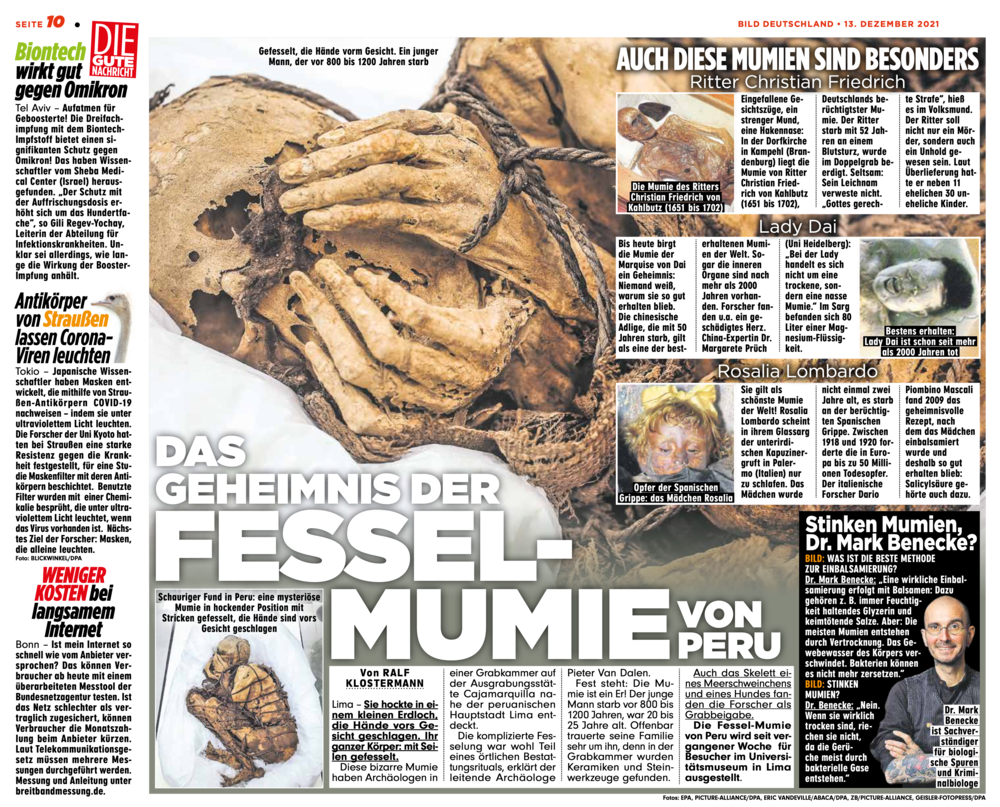
Das Geheimnis der Fesselmumie
von Peru

Mumien in Palermo
Buch

Lenins Leiche
Manche Tote leben länger
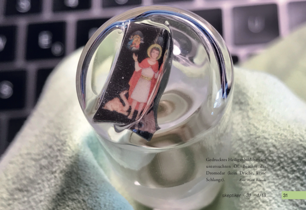
Heilige Tränen?
Skeptiker 2018

Neglect of the elderly
Forensic entomology cases and considerations

Severe post mortem damages
by ants on a human corpse
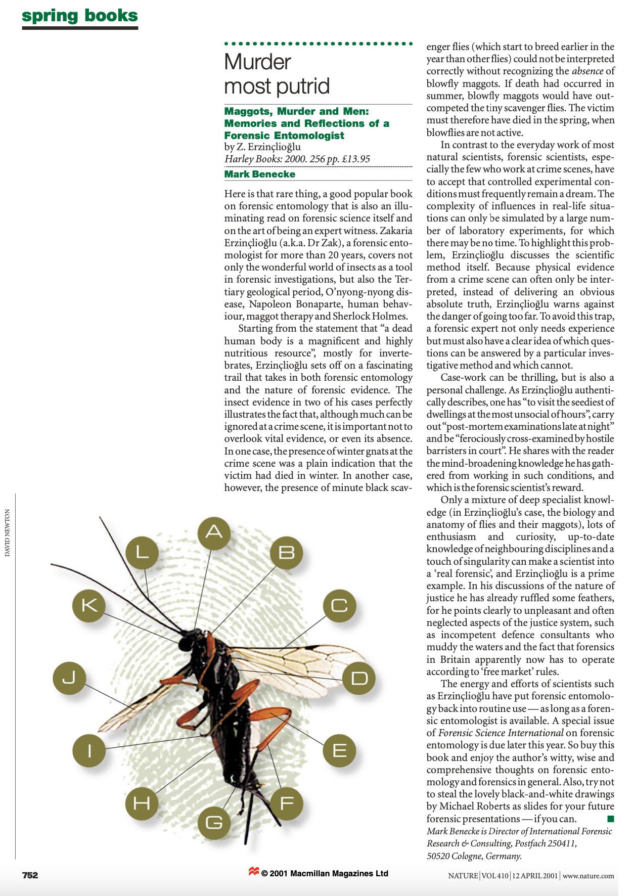
Murder most putrid
Memories & reflections of a forensic entomologist