Source: Forensic Science International (1998): 98(3): 157–168
The article as .pdf
Von Mark Benecke
Abstract
To permit quick identification of arthropods, random amplified polymorphic DNA typing (RAPD) was used to support classical morphological and medico-legal analysis of maggots on a human corpse. The method was employed to determine if maggots which were found on the inside of a body bag were identical (a) with maggots found on the outside of the bag, and (b) pupae found on the floor under the corpse. Pre-mixed RAPD reaction beads together with semiautomatic computer aided analysis of the PCR products are shown to discriminate between closely related necrophageous insect species (flies and beetles) found on corpses. From the 11 RAPD primers used, one alone was sufficient in resolving a practical forensic situation. This is the first report of a forensic application of RAPD DNA typing.
Keywords
Random amplified polymorphic DNA fingerprints, RAPD, Validation, Insects, Forensic entomology
1. Introduction
Recently, the use of arthropods in criminal forensic studies has become more recognized amongst forensic scientists, forensic pathologists, criminalists, jurists, and the public [1,2,31]. Subsets of insect and mite species were shown to be valuable tools in the investigation of post mortem intervals, even of badly decomposed corpses, child neglect, relocation of a body, identification of suspects, hygienical questions, and the explanation of two sets of postmortem lividities on one corpse [3–5]. Identification of the arthropod species found at the site of, or on a corpse, is essential but often very difficult if performed by morphological means. One reason being that the order of insects consists of more species than any other form of life which means that there are few experts for a whole insect family or insect order. Another reason for difficulty in insect determination are the minuscule morphological differences between species. In addition, the characteristics of immature larvae are sometimes extremely hard to determine. In the field of forensics these characteristics can become a focus of interest, especially in high profile cases where all other tools of criminalistics did not lead to final conclusions on the sequence of events (for example [2,4]).
Less than 40 scientists are actively engaged in the area of forensic arthropod examination [6] while only one works in a medico-legal institution. Due to the vast expertise required to determine (local) insect populations, it would be useful if the DNA laboratories of medico-legal institutes were able to support the forensic entomologists with DNA typing of forensic insect specimens but without disturbing their own routine work.
Here we report the use of a DNA typing technique — random amplified polymorphic DNA (RAPD) — as a quick and reliable tool for this special application.
We live in a world of insects with an immense species diversity (insects representing more than 80% of all species), therefore one of the main problems in typing forensic DNA of hexapodes, is a lack of suitable PCR primers for as many species as possible. Random amplified polymorphic DNA typing primers, in contrast to STR primers, are known to produce unique patterns for many animal and plant species. The primers are inexpensive, easy to store (dry at room temperature), and they produce highly informative and distinctive DNA profiles. On the other hand, the method is susceptible to changes in the reaction parameters including the type of thermocycler used. To overcome this, we used standardized PCR reaction beads (Ready-To-Go, Amersham Pharmacia Biotech) and validated the protocol so that the influences which might still mistakenly change RAPD DNA typing results were minimized.
2. Materials and methods
2.1. Specimens and DNA extraction
DNA was extracted from fresh adult and juvenile animals collected from decaying corpses (including the one actual case) delivered to the Institute for Forensic Medicine, Cologne University, Germany, and from dried specimens collected from the same source. Extraction was performed using overnight Proteinase K digest (DNAse free Proteinase K, Boehringer Mannheim) followed by phenol extraction without chloroform (TE saturated phenol, BRL Life Technologies). All specimens were washed in 70% ethanol prior to extraction. Whenever possible, maggots in their late developmental stages (i.e. with naturally empty guts) were used to prevent cross contamination with foreign DNA.
2.2. RAPD primers
Primers were synthesized for Amersham Pharmacia Biotech by Genosys Biotechnologies, Inc. and had the sequences given in Table 1.
2.3. Standardized reaction beads and amplification (PCR)
Premixed and dehydrated reaction beads delivered in 0.5 ml PCR tubes were used (Ready-To-Go RAPD Analysis Beads). The beads are composed of AmpliTaq DNA polymerase, Stoffel fragment, dNTPs, bovine serum albumin (BSA), reaction buffer, and an inert medium. They were stored in the dark at constant room temperature (258C).
Before PCR, beads were rehydrated with sterile deionized water, and 25 pM primer was added. Final concentrations in a total of 25 ml reaction mix: 1.5 units Taq, 10 mM Tris–HCl (pH 9.0), 50 mM KCl, 1.5 mM MgCl , 200 mM of each dNTPs and stabilizers 2 including BSA, 25 pM primer. DNA amount was varied around ,50 ng. PCR program: 45 cycles; preheat 958C, 5 min, denaturation: 958C, 1 min, annealing: 368C, 1 min, extension: 728C, 2 min. PCR was performed on two Perkin Elmer 2400 (one block each), one Biometra Uno (one block) and two Biometra Trio machines (three blocks each).
2.4. Electrophoresis and detection of RAPD PCR products
PCR products were separated on denaturing polyacrylamide gels on ALF express DNA sequencers (Amersham Pharmacia Biotech). Electrophoresis conditions: 5.5% PAG, time 500 min at 1200 V, 35 W. Reading interval between measurements of fluorescent light 2 s. Cy 5 labelling was excited by a non-moving beam of red laser light (2 mW) and detected by one single photo receptor per lane.
Samples amplified from the same DNA source were never loaded next to each other to avoid artificial (false) positives. For data recording and data processing, the ALF Manager 3.01 and the ALF Fragment Manager 2.1 software was used.
3. Results
3.1. Preliminary validation study
3.1.1. Influence of the PCR thermocycler on the reaction
Amplifying DNA extracted from one source three times each, different thermocyclers generally produced peaks at identical positions if all other reaction parameters were constant (reaction volume, reaction beads/mix, same experimentator) but the peaks were not necessarily of the same height and shape (Fig. 1). Major peaks remain at a height which exceeds all other peaks, and therefore only these major peaks should be looked at.
The ALF Fragment Manager allows the display of results not only as peaks (time scale horizontal) but also similar to the bands seen in silver-stained PAG (time scale vertical). Together with internal and external sizers, this display helps to easily detect the major peaks. An adequate threshold limit (10–40% of the height of the maximum peak, depending on the primer) can be set similar to the thresholds used to cut away so-called STR slippage peaks (reaction artifacts). A defect thermoblock (PE2400 2, Fig. 1) led to a total allele drop out.Well-to-well variation did not seem to have an obvious influence on the RAPD results.
3.1.2. Influence of the DNA amount
As in all PCR reactions, decrease of the total DNA amount under a certain level leads often to allele drop outs. In a range of 25 ng to 50 ng total DNA, allele drop outs could not be observed, except in a single case out of ca. 250 reactions (Fig. 2). In the regions of higher molecular PCR product (sizes exceeding 300 bp), only the major peaks were still visible which may be desirable in the prevention of non-informative small peaks.
The cut-off limit depends on the primer used and should be set as high as possible. An average concentration of 50 ng was sufficient in producing a coherent profile. There were no relevant differences in the DNA profiles within a range of 20 and 60 ng total DNA in a 25-ml total volume PCR reaction.
3.1.3. Reproduction of amplification results
In all maggots found in a real case (see next paragraph), the RAPD DNA pattern was identical for all DNA extracts amplified on the same thermocycler (Biometra Trio), even if the PCR was performed on different days and with two different total concentrations of DNA each (Fig. 3). Again, peak shape did vary but peak height was constant between the different amplificates. A major problem we observed were ‘splitting peaks’ which means that broad peaks tend to split up in several peaks or to produce more than one top of the peak, or vice versa that two or more peaks unify to one. Frequently, this problem can be overcome by switching the electrophoretic display from peak view to a silver-stain-like banding pattern view.
3.1.4. Influence of the primer
Most of the primers produced hundreds of bands of which only the most intense (highest peaks, usually .50% of maximum intensity) were scored as being relevant and useful. The smaller peaks varied too much and did not lead to further information. Any two to three out of our set of 11 primers produced useful results. For actual casework, we start the analysis with REP1R and Primer 5 but switch to other primers if the number of high peaks is not sufficient.
3.2. A case: identity of maggots found on the outside and the inside of a body bag
In October 1997, a body in severe decay was analyzed for insect evidence to determine the post mortem interval. Hundreds of maggots of an average size of 9 mm were found on the corpse and on the outside of the closed body bag. It is known that maggots squeeze themselves through tiny holes, e.g. to find a place for pupation [4], the maggots outside might have come through holes from the inside. Alternatively, a second
oviposition of another species might have taken place after the body was put into the bag.
Furthermore, on the floor beneath the corpse, pupae were found which could not be directly related to any specific body in the morgue, i.e. they may have fallen down from several unknown corpses. Since different fly species develop within different times, post mortem interval estimation is only possible if the insect species is known and can thus be used as a time since death indicator. The species of maggots — especially of young ones — is hard to identify, and for this reason we searched for a quick, inexpensive and reliable DNA test which helps in acute forensic situations in which, for whatever reason, no classical insect analysis can be performed (e.g. no entomologist available, urgent case which does not allow breeding of the insects).
RAPD beads are known to be a help in cases dealing with a variety of species. Furthermore, the beads can be stored at room temperature, are both quick and easy to use, and are composed of defined amounts of reagents.
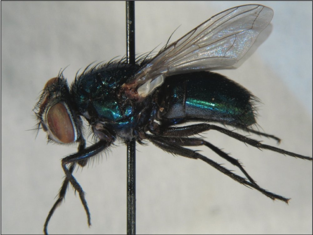
In our case, we used maggots from the inside and the outside of the body bag which were later determined as ‘green bottle’ blow flies Lucilia spec. (Diptera: Calliphoridae) and compared them to dry stored adults of ‘blue bottle’ blow flies Calliphora erythrocephala (Diptera: Calliphoridae) from another case, and adult silphid carrion beetles (Coleoptera: Silphidae).
As shown in Fig. 4, the profiles of four different maggot individuals from the inside of the body bag are similar over the whole stretch of 460 min (in our pictures, longer PCR products are on the right, shorter ones on the left). However, some shapes and heights of some peaks differ in the way described above. Using only the highest peaks produced by the primers REP1R and Primer 5, it was possible to distinguish between different species. It was also found that the pupa was not just a different (later) stage of one of the maggots from the actual corpse. Except for two major bands which might be a distinctive sign of blow flies, there was no similarity between the DNA profiles of the two fly species and between the profiles produced by the different primers. The DNA profile of the beetle (exterior control) did not show any similarities to the blow fly profiles.
4. Discussion
Since the first reports on the discriminating power of RAPD DNA typing in insect-related ecological questions [7–9] an increasing number of scientists made use of the fast and highly informative DNA typing method. Nowadays, not only vertebrates (commercial breeding [10], research on endangered species [11], inbreeding in wildlife species [12]) but also several insects (beetles: [13–15]; flies: [16,17], dragonflies: [7,18]) and a variety of bacterial, animal and plant species [19–24] were successfully DNA typed with RAPDs. From the beginning it was clear that the method has its limitations since different reaction parameters can lead to different DNA typing results [25,26]. At the same time, standardization and mathematical approaches led to significant improvement of the method [27–29], processes which are well known in medico-legal DNA typing [30].
Although it is thought that the model/brand of thermocyclers and their intrinsic temperature profiles (especially the rate of heating and cooling with or without electronic Peltier thermoregulation) used in the polymerase chain reaction is one of the main factors producing differences in the RAPD profiles of identical specimens, we could find enough similarities in the DNA profiles under our highly standardized conditions (reaction beads). Results that did differ significantly were due to defective thermocyclers or blocks (e.g. Fig. 1; PE 2400 block 2). The total amount of DNA in the PCR is relevant but produces identical results within a range of concentrations. This was also found in an earlier validation study [30] where all concentrations in a range of 1 ng to 10 ng total DNA produced reproducible results with another set of primers (Operon primers).
In forensic practice, the shapes and heights of the RAPD peaks differ from each other (Fig. 3). A detailed analysis of the peaks, a restriction to certain areas of the peak profile and especially the use of a cut-off value for all peaks helps to make use of the quick and multifunctional RAPD method. This means that RAPD beads are a helpful tool especially for investigations which cannot be performed by any other means; for forensic purposes, a set of at least six primers should be used to establish similarity coefficiencies.
In urgent cases, and where maggots cannot be matured in the lab to determine the species from the adult, RAPDs may also be considered as a helpful tool. The advantage of the high information density of RAPDs (many variable bands) can be difficult to handle. To lower the information density obtained out of RAPD profiles, it is helpful to restrict oneself to certain areas of the RAPD profile and to use a cut-off value. This reduces the number of bands to an amount which can be dealt with. For a comparison of the advantages and disadvantages of the method, see Table 2.
We are aware that in medico-legal matters, RAPD results may only be reported for so-called exclusions (where two specimens are definitely proven to be different) since an inclusion (where two specimens are shown to be similar or directly related) might induce the question of the likelihood of finding the same RAPD pattern by chance in any other animal. The question of similarity is hard to answer without databases which might not be established due to the high number of forensic insect species, because of the non-species specific properties of the primers, and the possible noncompatibility of RAPD results between laboratories using different PCR parameters. A pure identification of insects is not possible at present, and it would be worth determining a universal scoring system for major peaks and threshold limits.
Our experiments and those of others [23] show that RAPD results can be transferred from pure research to practical applications, and that they are safe enough to allow direct therapeutic decisions (secondary infections, e.g. in AIDS patients), breeding supervision (e.g. to check for contaminations in bacteria cultures) and forensic investigations (as shown here). In forensics, especially the rapid identification of maggots which are often hard to determine by morphological features might be an invaluable tool in urgent cases where maturing of insects is not an option because of a lack of time. Furthermore, maggots from different ovipositional events can be distinguished.
Acknowledgements
The following individuals had a part in conducting the study presented here: Yvonne Jacobi, Stefan Tomiuk, Britta Jenke, Bertram Weiss (University of Cologne, Germany), Dirk Lehmann (University Osnabru¨ck, Germany), Heike Hadrys (Yale University, New Haven, USA), Peter Wiesner (Amersham Pharmacia Biotech Europe/LION AG, Heidelberg, Germany), Madelaine Nash (Milton Helpern Library for Legal Medicine/New York University Medical School), and Emma Tibbo (Office of Chief Medical Examiner, NYC). All experiments were performed at the Institute for Legal Medicine and the Institute of Biochemistry of the University of Cologne, Germany. In 1997, the author was supported by a Fortu¨ne scholarship.
References
[1] M. Benecke, Rechtsmedizinisch angewandte kerb- und spinnentierkundliche Begutachtungen in Europa: eine kurze Übersicht über Ursprünge und den aktuellen Stand der Forschung. (Use of insect evidence in history and in Europe’s forensic medicine — a short survey), Rechtsmedizin 8 (1998) 153–155.
[2] M. Benecke, B. Seifert, Insect evidence as a crucial clue in a high profile murder case (in preparation).
[3] M. Benecke, Zur insektenkundlichen Begutachtung in Faulleichenfällen (Expert insect identification in cases of decomposed bodies), Arch. Kriminol. 198 (1996) 99–109.
[4] M. Benecke, Six forensic entomology cases: description and commentary, J. Forensic Sci. 43 (1998) 797–805.
[5] W.D. Lord, Case histories of the use of insects in investigations, in: P.E. Catts, N.H. Haskell (Eds.), Entomology and Death. A Procedural Guide, Joyce’s Print Shop, Clemson, 1990, pp. 9–37.
[6] http: / /www.uio.no/|mostarke/ forens ent / forensic entomologists.html
[7] H. Hadrys, B. Schierwater, S.L. Dellaporta, R. DeSalle, L.W. Buss, Determination of paternity in dragonflies by random amplified polymorphic DNA fingerprinting, Mol. Ecol. 2 (1993) 79–87.
[8] H. Hadrys, M. Balick, B. Schierwater, Applications of random amplified polymorphic DNA (RAPD) in molecular ecology, Mol. Ecol. 1 (1992) 55–63.
[9] N.J. Gawel, A.C. Bartlett, Characterization of differences between whiteflies using RAPDPCR, Insect Mol. Biol. 2 (1993) 33–38.
[10] S.K. Semenova, A.L. Tilenko, V.A. Vasil’ev, M.L. Prosniak, A.A. Sevastíanova, A.P. Ryskov, Ispolízovanie polymorfnykh markerocv DNK dlia differentsiatsii porod kur razlichnogo projskhozhdeniia, Genetika 32 (1996) 795–803.
[11] J.A. Nasser, R.M. Goto, D.B. Ledig, R.C. Fleischer, M.M. Miller, RAPD analysis reveals low genetic variability in the endangered light-footed clapper rail, Mol. Ecol. 5 (1996) 463–472.
[12] P. Shankaranarayanan, M. Banerjee, R.K. Kacker, R.K. Aggarwal, L. Singh, Genetic variation in Asiatic lions and Indian tigers, Electrophoresis 18 (1997) 1693–1700.
[13] R.J. Brown, C.A. Malcolm, P.L. Mason, R.A. Nichols, Genetic differentiation between and within strains of the saw-toothed beetle, Oryzaephilus surinamensis (Coleoptera: Silvanidae) at RAPD loci, Insect Mol. Biol. 6 (1997) 285–289.
[14] M.C. Carter, J.L. Robertson, R.A. Haack, R.K. Lawrence, I.L. Hayes, Genetic relatedness between North American populations of Tomicus piniperda (Coleoptera: Scolytidae), J. Econ. Entomol. 89 (1996) 1345–1353.
[15] B.L. Apostol, W.C. Black, P. Reiter, B.R. Miller, Population genetics with RAPD-PCR markers: the breeding structure of Aedes aegypti in Puerto Rico, Heredity 76 (1996) 325–334.
[16] J. Stevens, R. Wail, Species, sub-species and hybrid populations of the blowflies Lucilia cuprina and Lucilia sericata (Diptera: Calliphoridae), Proc. R. Soc. London Ser. B Biol. Sci. 263 (1996) 1335–1341.
[17] A. Sonvico, F. Manso, L.A. Quesada-Allue, Discrimination between the immature stages of Ceratitis capitata and Anstrepha fraterculus (Diptera: Tephritidae) populations by random amplified polymorphic DNA polymerase chain reaction, J. Econ. Entomol. 89 (1996) 1208–1212.
[18] R.E. Hooper, M.T. Siva-Jothy, Last male sperm precedence in a damselfly demonstrated by RAPD profiling, Mol. Ecol. 5 (1996) 449–452.
[19] M. Benecke, DNA typing in today’s forensic medicine and criminal investigations. A current survey, Naturwissenschaften 84 (1997) 181–188.
[20] J.P. Bielawski, D.E. Pumo, Randomly amplified polymorphic DNA (RAPD) analysis of Atlantic Coast striped bass, Heredity 78 (1997) 32–40.
[21] I. Sivolap, I.A. Balashova, L.P. Troshin, Issledovanie geneticheskogo polimorfizma vinograda pripomoshchi RAPD-analiza, Tsitologiia i Genetica 30 (1996) 33–37.
[22] A. Ender, K. Schwenk, T. Stadler, B. Streit, B. Schierwater, RAPD identification of microsatellites in Daphnia, Mol. Ecol. 5 (1996) 437–441.
[23] S.C. Chen, A.G. Brownlee, T.C. Sorrell, P. Ruma, G. Nimmo, Identification by random amplification of polymorphic DNA of a common molecular type of Cryptococcus neoformans var. neoformans in patients
with AIDS or other immunosuppressive conditions, J. Infect. Dis. 173 (1996) 754–758.
[24] R.E. Veilleux, L.Y. Shen, M.M. Paz, Analysis of the genetic composition of anther-derived potato by randomly amplified polymorphic DNA and simple sequence repeats, Genome 38 (1995) 1153–1162.
[25] W.C. Black, PCR with arbitrary primers: approach with care, Insect Mol. Biol. 2 (1993) 1–6.
[26] B. Schierwater, A. Ender, Different thermostable DNA polymerases may amplify different RAPD products, Nucl. Acids Res. 21 (1993) 4647–4648.
[27] W.E. Lamboy, Computing genetic similarity coefficients from RAPD data: the effects of PCR artifacts, PCR Methods Appl. 4 (1994) 31–37.
[28] W.E. Lamboy, Computing genetic similarity coefficients from RAPD data: correcting for the effects of PCR artifacts caused by variations in experimental conditions, PCR Methods Appl. 4 (1994) 38–43.
[29] Q. He, M.K.Viljanen, J. Mertsola, Effects of thermocyclers and primers on the reproducibility of banding patterns in randomly amplified polymorphic DNA analysis, Mol. Cell. Probes 8 (1994) 155–160.
[30] M. Benecke, Bestimmung inter- und intraartlicher Unterschiede zwischen verschiedenen Nematodenpopulationen mittels genetischer Fingerprintverfahren. Diploma thesis, Faculty of Natural Sciences, University of Cologne, Germany. Verlag der Schillerbuchhandlung, Köln, 1995.
[31] G.S. Anderson, The use of insects to determine time of decapitation: a case-study from British Columbia, J. Forensic Sci 42 (1997) 947–950.

Damages by ants
Corpses colonization

Neglect of the elderly
Forensic entomology
Flies and ant lesions
Case report from Italy

When DNA does not help
Sexual assault and murder

Glauben Sie an die Besserung eines Menschen?
Reizverschluss 2017
DXS10011
A powerful marker for forensic purposes

Six forensic entomology cases
Description & commentary

Asian Y-STR
Haplotype reference database